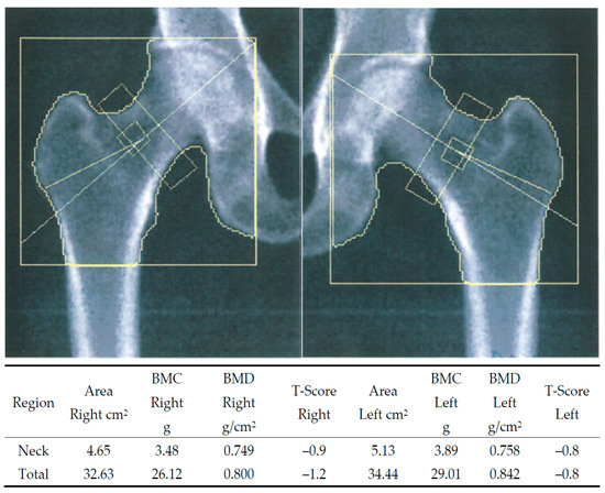
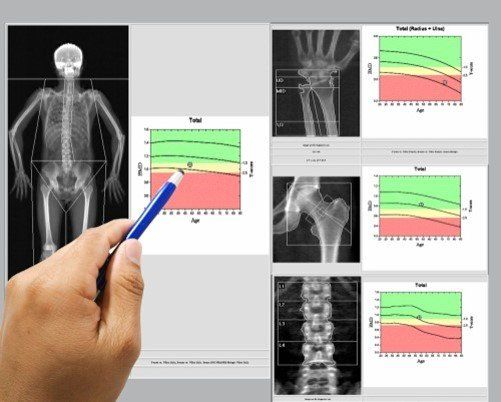
Bilateral hip DXA scan image from a 59-year-old post-menopausal woman.
-
By A Mystery Man Writer
-
-
4.7(206)

Product Description
Download scientific diagram | Bilateral hip DXA scan image from a 59-year-old post-menopausal woman. The dominant arm did not match, but dominant leg did. The T-score for the lumbar spine was normal. If the patient had only had her left hip examined in accordance with the dominant arm, the conclusion would have been normal bone mineral density (BMD). Having both hips examined instead led to the conclusion of low bone density (LBD). from publication: Dual-energy X-ray Absorptiometry of Both Hips Helps Appropriate Diagnosis of Low Bone Mineral Density and Osteoporosis | Controversy still remains regarding the use of bilateral hip scanning when bone mineral density (BMD) is measured, and bilateral hip scanning is not mandatory in international guidelines for screening of osteoporosis. BMD of both hips and the lumbar spine was analyzed in 133 | Hip, Dual-Energy X-ray Absorptiometry and Bone Mineral Density | ResearchGate, the professional network for scientists.
Diagnostics, Free Full-Text

2006 Abstracts: Twenty‐Eighth Annual Meeting of the American Society for Bone and Mineral Research: Pennsylvania Convention Convention Center Philadelphia, Pennsylvania, USA, September 15–19, 2006 - 2006 - Journal of Bone and Mineral Research - Wiley

Imaging in Osteoporosis and Paget's disease

a Sample images of lateral vertebral fracture assessment by DXA in

Osteoporosis Imaging: State of the Art and Advanced Imaging

Evolutionary roots of the risk of hip fracture in humans

Best Practice Recommendations for DXA Scans and Reports
Osteoporosis Workup: Approach Considerations, Laboratory Studies, Biochemical Markers of Bone Turnover
Medical Science Monitor Current Applications and Selected Technical Details of Dual-Energy X-Ray Absorptiometry - Article abstract #930839


:max_bytes(150000):strip_icc()/Health-GettyImages-1370512518-0f661134438c4aad84b4030eddea17d8.jpg)